 en Tendinopatía Rotuliana. Estudio a 10 años (ENG)")
CLINICAL RESULTS AFTER ULTRASOUND-GUIDED GALVANIC ELECTROLYSIS TECHNIQUE (USGET) AND ECCENTRIC EXERCISE IN THE TREATMENT OF PATELLAR TENDINOPATHY
 Abat F, et al.
© Springer-Verlag Berlin Heidelberg 2014
Introduction
Patellar tendinopathy or jumper’s knee is a frequent condition that most commonly affects the tendon’s origin on the inferior pole of the patella [2, 4, 10]. Once considered an inflammatory condition, it is currently considered a degenerative process due to the presence of myxoid degeneration, the disruption of the collagen fibres and signs of hypoxia in tenocytes and resident macrophages [6, 17].
The overall prevalence of patellar tendinopathy is around 14 % in the sports population [3, 16], but may be as high as 40 % in highly demanding athletes [8]. The tendon’s overuse in sports that involve running, jumping or rapid change in direction is considered the main risk factor for developing the said condition [16].
Current treatment options include eccentric training [15, 18, 29], open or arthroscopic surgery, extracorporeal shockwave therapy [25], ultrasound (US)-guided sclerosis [12], non-steroidal anti-inflammatory drugs, platelet-rich plasma injection [30] and aprotinin [1]. These studies have also suggested that, in general, patients with a worse functional status before treatment obtain inferior final outcomes. However, due to the limited evidence-based therapies, there are still several controversies regarding the real efficacy of these treatment modalities [1].
Ultrasound Guided Galvanic Electrolysis (USGET) treatment is a pioneering US-guided technique developed by one of the authors. It leads to a non-thermal electrochemical ablation through a cathodic flow directly at the clinical focus of degeneration. USGET causes an organic reaction leading to a highly localized inflammation, exclusively at the region of treatment that conduces to a rapid regeneration of the injured tendon [26].
The present study provides the first analysis of the results of USGET in the treatment of patellar tendinopathy at 10 years follow-up. This study could be clinically relevant given the lack of effective techniques in the treatment of patellar tendinopathy.
The aim of this study was to investigate the outcome of the USGET in terms of pain, function and the return to the previous level of activity in patients with patellar tendinopathy. The mean follow-up of 10 years provides information on safety and the rate of recurrence. The main hypothesis was that USGET would quickly improve the outcome in patients with patellar tendinopathy and that this improvement would be maintained over a long period of time. The second hypothesis was that good outcomes would be obtained regardless of the initial degree of functional impairment. It was also hypothesized that the patients would be restored to their pre-injury activity level.
Materials and methods
From January 2002 to October 2002, 41 patients with patellar tendinopathy were included in the investigation. Demographic data and patient information (age, gender, affected and dominant side, kind of sport or activity level) were recorded.
The inclusion criteria were a history of patellar tendon pain, tenderness upon palpation, functional limitation directly related to the studied tendon and sonographic confirmation of tendon degeneration. A tendon injury located at the inferior pole of the patella was considered a requisite. Other inclusion criteria were more than 4 weeks of symptoms and an age of \60 years old. Patients were classified according to Blazina’s scale [22]. Exclusion criteria were pain at the proximal pole of the patella (frequently included in jumper’s knee), chronic articular dis- ease, a concomitant knee pathology, contraindications to the USGET and the concomitant administration of certain drugs (at least 2 weeks before receiving treatment). The inclusion and exclusion criteria are summarized in Table 1 (see article).
Ultrasound examination
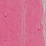
All the patients went through an exhaustive US examination of the tendon and adjacent structures using a high- resolution greyscale US (Fig. 1) with Doppler power and linear multi-frequency probe (6–15 MHz). The injured and the contralateral knees were studied in all patient. The US efficacy for the proper diagnosis of patellar tendinopathy was previously reported [11, 36, 37].

Ultrasound Guided Galvanic Electrolysis (USGET) protocol
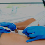
The USGET was applied using a specifically developed medically device which produces modulated galvanic electricity through the negative electrode cathodic flow. This is applied using a modified electrosurgical scalpel that uses acupuncture needles (0.3 mm in diameter) with different lengths. The intensity can be adjusted by changing the duration or the milliamps of the device. Conversely, the polarity of the machine is fixed (i.e. only the cathodic flow is usable). During the procedure, performed by the same experienced operator, the patients are supine so as to minimize any potential vagal reaction. Isopropyl alcohol was used to prepare the skin despite the bacteriostatic action of theUSGET. Polyvidone iodine was avoided to prevent a tattoo effect of the cathodic flow.
Finally, three US-guided precise punctures at 3 mA were performed until a complete debridement of the treated area was obtained. The debridement was assessed with the sonographic images. After the first USGET treatment, the patients underwent consecutive sessions ofUSGET every 2 weeks and 2 weekly sessions of an eccentric exercise training using the resistance isoinertial leg-press machine (YoYoTM Technology AB, Stockholm, Sweden).
Eccentric exercises were performed in three sets of ten repetitions twice a week in order to obtain maturation of collagen fibres [24, 31]. Each repetition was performed with the concentric phase with both extremities, whereas the eccentric phase was only performed with the affected limb at a maximum of 60° of knee flexion. Patients received USGET treatment up to a maximum of ten sessions. The treatment finished either when the patients were symptom free or if there was no improvement in terms of pain or function after those ten sessions.
Treatment evaluation
All the patients were evaluated before treatment and prospectively when their treatments were finished (at the third month), at 2-year, at 5-year and at 10-year follow-up.
The primary outcome measure was knee function using the Victorian Institute of Sport Assessment–Patella (VISA-P) score, a specific validated questionnaire to quantify pain and knee function and ability to play sport in patients with patellar tendinopathy [9, 34]. The VISA-P score ranged from a maximum of 100 in asymptomatic patients to the theoretical minimum of 0. The authors of the score suggested that a score between 80 and 100 points might be considered as the optimal outcome category. Functional evaluation was further assessed with Blazina’s classification [22]. This classification categorizes the symptomatic patients as in phase I (pain only after activity), phase II (discomfort during activity), phase III (pain during activity that interferes with participation) and phase IV (complete tendon disruption). The Tegner score was also used to assess the influence of the treatment in terms of restoring the previous sports activity level. All the written questionnaires were personally filled out by all patient before treatment, at the end of the treatment (at 3-month) and at the 2-year follow-up. The questionnaires corresponding to the 5- and 10-year follow-up evaluations were all filled out through a telephone interview. Patient satisfaction was measured according to the Roles and Maudsley score [23]. In this score, patients are classified as Excellent (no pain, full movement and full activity), Good (occasional dis- comfort, full movement and full activity), Fair (some discomfort after prolonged activity) or Poor (pain limiting activities).
All those patients that scored\50 points with the VISA- P questionnaire at baseline were denominated Group 1, whereas the remaining patients scoring equal to or higher than 50 points were denominated Group 2. This classification allows to display the results in different degrees of injury of the patellar tendon: more (VISA-P \ 50 points) or less affected (VISA-P [ 50 points).
The Clinical Research Ethics Committee of ICATME- Institut Universitari Dexeus, University of Barcelona, approved the study (09/06/0049). All the patients signed informed consent to participate in the study as well as for the evaluation and publication of their results.
Statistical analysis
Categorical variables are presented as number of cases and percentages. Continuous variables are presented as mean ± SD (range). The relationships between categorical variables were described using contingency tables, and inference was studied using the chi-square test or Fisher’s exact test. The relation between the VISA-P score and dichotomous variables was assessed using the Mann– Whitney test, showing the median value. Analysis of variance (ANOVA) was used to compare the evolution between groups. Statistical significance was set at 0.05 two-sided. Statistical analysis was performed using SPSS 19 (SPSS Inc., Chicago, IL, USA).
Results
One patient was lost during the first 3 months of follow-up. The remaining 40 patients were available at the 3-month and at the 2-year evaluations. At the 5-year evaluation, another three patients were lost (37 patients available, 90.2 % of the cases) and another three patients at the 10-year assessment (34 patients available, 82.9 % of the cases).
Patient description
Twenty-one patients (52.5 %) were included in Group 1 and the remaining 19 (47.5 %) in Group 2. Both groups were comparable in terms of age, gender, side and functional scores at baseline. Sports involvement is summarized in Table 3 (see article). No relation (n.s.) between the injured tendon and the dominant extremity, the type of sport, the age of the patient and gender, and the VISA-P values obtained after the treatments was observed.
The mean duration of symptoms prior to the treatment was 69.4 ± 65.6 weeks (range 4–288 weeks). The athletes were off sports activities due to their patellar tendinopathy for a mean time of 40.6 ± 50.9 weeks (range 0–192 weeks). Treatment duration averaged 7.5 ± 2.6 weeks (range 1–10 weeks), and the patients required a mean of 4.1 ± 2.6 USGET procedures (range 1–10). According to Blazina’s classification, one patient (2.5 %) was of stage I at baseline, seven patients (17.5 %) stage II and the remaining 32 patients (80 %) stage III. At the 3-month evaluation, once all the treatments were finished, five patients (12.5 %) were classified as of stage I and six patients (15 %) stage II. All the remaining 30 cases (72.5 %) were considered completely cured (less than Blazina’s stage I). At the 2-year follow-up evaluation, 31 cases (77.5 %) were asymptomatic (less than Blazina’s stage I) and nine (22.5 %) were in stage I. Analysis of the patients using the Blazina’s classification remained unchanged throughout the remaining follow-up evaluations of the period studied (n.s.).
Clinical outcomes over time
The VISA-Pand Tegner scores before treatment, at 3 months and at 2, 5 and 10 years of follow-up are summarized in Table 4. Group 1 improved by 45.8 points (p \ 0.001) at 3 months to obtain a mean VISA-P score of 78.9 ± 14.4. In Group 2, the mean improvement in VISA- P score at 3 months was 15.6 points at 3 months (p \ 0.001). The Tegner level did not drop over the 10 years of the study period, and no differences between the intermediate evaluations (n.s.) were observed either.

According to the Roles and Maudsley score, patient satisfaction at 3 months of follow-up was considered Excellent in 32 cases (80 %), Good in seven cases (17.5 %) and Fair in one case (2.5 %). These values persisted without significant differences throughout the period studied. No recurrences, adverse episodes or any additional modality of treatments were reported after the 10 years of follow-up.
At the 3-month follow-up evaluation, 32 (80 %) patients restored their previous activity level according to Tegner scale (n.s.). In eight patients (20 %), there was a decrease in only one single level on the same scale. These values were maintained over the remaining period studied (n.s.).
Discussion
Treatment with USGET in combination with eccentric exercises has been shown to effectively improve the symptoms of patellar tendinopathy quickly and steadily for at least 10 years. It confirmed the first hypothesis. This improvement in patients that had different severities of VISA-P scores at baseline was equally obtained in terms of symptomatology, knee function and return to sports activity, which is also in concordance with the second hypothesis. The results observed in the first study reporting on the clinical use of USGET are encouraging [26]. Its effects are based on a local and non-thermal electrochemical therapy that induces a localized short inflammatory response through an electrolytic reaction produced by a cathodic flow. Consequently, this causes an organic reaction leading to the regeneration of the injured tendon [26].
Conservative treatment was traditionally considered the first option of treatment of tendinopathies. Many different techniques were used [1, 8], such as modification of activity, eccentric physical training, patellar straps, cold and heat compression transfriction massage and stretching for quadriceps, hamstrings and patellar tendons. Despite some good results reported with eccentric programmes [18, 28], it is still unclear as to the more effective exercise protocol, its frequency, load and dosage. While Zwerver et al. [37], in a recent randomized clinical trial, concluded that no benefit came of extracorporeal high-energy shock- wave therapy during competition, Rompe et al. [25] reported, at 4-month follow-up, that eccentric loading alone was less effective when compared with a combination of eccentric loading and repetitive low-energy shock- wave treatment. Similarly, low-intensity US is not currently considered a reliable method for the treatment of patellar tendinopathy [14, 15, 35].
Different injection treatments for patellar tendinopathy have been proposed. While some studies on the effect of dry needling, autologous blood and high volume have been put forward as providing functional improvements, steroid treatment has shown a relapse of symptoms after few months, not to mention the deleterious effect on the tendon histology [32]. Recent investigations have observed slightly better outcomes after treatment with platelet-rich plasma injections in association with an eccentric training programme than an eccentric training programme alone in short-term studies [7, 30, 32]. Some authors had initially reported pain relief after sclerosing injections of polidocanol [10], but recent studies have shown contradictory results [33]. Hoksrud et al. reported their results with US- guided sclerosis of neovessels in 29 patients with 44 months of follow-up [12] and in 101 patients with 24 months of follow-up [13]. The patients needed several injections over 8 months of treatment, and only a moderate improvement in knee function was observed. One-third of their patients obtained a VISA-P score \50 points, and only few patients were completely cured. Conversely, in the present investigation with short- and long-term reported outcomes, even the patients with lowest VISA-P score (\50 points) at baseline significantly improved to around 80 points at 3 months and to around 90 points at 10 years. These final outcomes were comparable with those obtained by the patients with better VISA-P scores before treatment. This is of considerable relevance because the professional sports patients included in this series started from lower VISA-P values and they still obtained excellent scores. Overall, 80 % (n = 32) of the treated patients returned to the same level of sports activity at 3 months, and the remaining eight patients only decreased a single level in the Tegner score.
Regarding surgical treatment of patellar tendinopathy, some open [5, 21] and arthroscopic [5, 20, 27] techniques have also been recommended when conservative treatment fails. However, surgery usually provides unpredictable and inconsistent results [4, 15], which is often no more effective than an isolated eccentric exercise programme [2], and it does not allow the athletes to resume their previous sports at the same level, at least within the first year of treatment [19].
The main strengths of the current study are that, as far as we know, it is the first investigation reporting on any treatment modality for patellar tendinopathy over the course of 10 years. Few patients were lost during this long follow-up period. In addition, it is also the first study reporting on the clinical outcome using the USGET in the treatment of tendinopathy at long term follow-up. The promising results obtained with the USGET procedure showed excellent functional results assessed with the VISA-P score as well as with the Blazina’s classification in around 80 % of the patients at 3 months and over 90 % at 10 years. It also allowed a full recovery to the previous activity level in most patients. This outcome’s improvement with the use of USGET in the treatment of patellar tendinopathy was achieved after a short period of time (mean 7.5 weeks) and with a few number of treatment sessions (mean 4.1 USGET treatments).
Besides the low sample size, one of the most relevant limitations of the current study is the lack of a control group. Comparison with a placebo-treated group of patients would have made for much stronger conclusions. However, most of our patients were professional or semi- professional athletes referred by other physicians after failure of conservative therapy. It seems highly unlikely that this sort of patients would be willing to accept placebo treatment for a long enough period. Another weak- ness might be that the combination of treatment with eccentric exercises might have positively affected the results attributed to the USGET. Although this could more logically affect the results during the first months of follow-up, it does not seem that it should have had any influence in the long-term results. Regardless of the aforementioned limitations, this study provides the first analysis of the EPIÒ technique on the treatment of patellar tendinopathy, with promising results after a long follow-up period.
The clinical relevance of the reported results was that USGET brought about a major improvement in pain and function in comparison with the so far known techniques and offers a good treatment option in patellar tendinopathy.
Conclusion
Treatment with Ultrasound Guided Galvanic Electrolysis (USGET) and eccentric exercises in patellar tendinopathy resulted in a great improvement in knee function and a rapid return to the previous level of activity after few sessions. The procedure has proved to be safe with no recurrences on a long-term basis.
References
- Andres BM, Murrell GA (2008) Treatment of tendinopathy: what works, what does not, and what is on the horizon. Clin Orthop Relat Res 466:1539–1554
- Bahr R, Fossan B, Loken S, Engebretsen L (2006) Surgical treatment compared with eccentric training for patellar tendin- opathy (jumper’s knee): a randomized, controlled trial. J Bone Jt Surg Am 88:1689–1698.
- Cannell LJ, Taunton JE, Clement DB, Smith C, Khan KM (2001) A randomised clinical trial of the efficacy of drop squats or leg extension/leg curl exercises to treat clinically diagnosed jumper’s knee in athletes: pilot study. Br J Sports Med 35:60–64
- Coleman BD, Khan KM, Maffulli N, Cook JL, Wark JD (2000) Studies of surgical outcome after patellar tendinopathy: clinical significance of methodological deficiencies and guidelines for future studies. Victorian Institute of Sport Tendon Study Group. Scand J Med Sci Sports 10:2–11
- Coleman BD, Khan KM, Kiss ZS, Bartlett J, Young DA, Wark JD (2000) Open and arthroscopic patellar tenotomy for chronic patellar tendinopathy: a retrospective outcome study, Victorian Institute of Sport Tendon Study Group. Am J Sports Med 28:183–190
- Coombes BK, Bisset L, Vicenzino B (2010) Efficacy and safety of corticosteroid injections and other injections for management of tendinopathy: a systematic review of randomised controlled trials. Lancet 376:1751–1767
- Filardo G, Kon E, Della Villa S, Vincentelli F, Fornasari PM, Marcacci M (2010) Use of platelet-rich plasma for the treatment of refractory jumper’s knee. Int Orthop 34:909–915
- Fredberg U, Bolvig L, Andersen NT (2008) Prophylactic training in asymptomatic soccer players with ultrasonographic abnor- malities in Achilles and patellar tendons: the Danish Super League Study. Am J Sports Med 36:451–460
- Hernandez-Sanchez S, Hidalgo MD, Gomez A (2011) Cross- cultural adaptation of VISA-P score for patellar tendinopathy in Spanish population. J Orthop Sports Phys Ther 41:581–591
- Hoksrud A, Ohberg L, Alfredson H, Bahr R (2006) Ultrasound- guided sclerosis of neovessels in painful chronic patellar ten- dinopathy: a randomized controlled trial. Am J Sports Med 34:1738–1746
- Hoksrud A, Ohberg L, Alfredson H, Bahr R (2008) Color Doppler ultrasound findings in patellar tendinopathy (jumper’s knee). Am J Sports Med 36:1813–1820
- Hoksrud A, Bahr R (2011) Ultrasound-guided sclerosing treat- ment in patients with patellar tendinopathy (jumper’s knee). 44-Month follow-up. Am J Sports Med 39:2377–2380
- Hoksrud A, Torgalsen T, Harstad H, Haugen S, Andersen TE, Risberg MA, Bahr R (2012) Ultrasound-guided sclerosis of neovessels in patellar tendinopathy: a prospective study of 101 patients. Am J Sports Med 40:542–547
- Khanna A, Nelmes RT, Gougoulias N, Maffulli N, Gray J (2009) The effects of LIPUS on soft-tissue healing: a review of litera- ture. Br Med Bull 89:169–182
- Larsson ME, Ka ̈ll I, Nilsson-Helander K (2012) Treatment of patellar tendinopathy—a systematic review of randomized con- trolled trials. Knee Surg Sports Traumatol Arthrosc 20: 1632–1646
- Lian OB, Engebretsen L, Bahr R (2005) Prevalence of jumper’s knee among elite athletes from different sports: a cross-sectional study. Am J Sports Med 33:561–567
- Maffulli N, Khan KM, Puddu G (1998) Overuse tendon condi- tions: time to change a confusing terminology. Arthroscopy 14:840–843
- Malliaras P, Barton CJ, Reeves ND, Langberg H (2013) Achilles and patellar tendinopathy loading programmes: a systematic review comparing clinical outcomes and identifying potential mechanisms for effectiveness. Sports Med 43:267–286
- Panni AS, Tartarone M, Mafulli N (2000) Patellar tendinopathy in athletes. Outcome of nonoperative and operative management. Am J Sports Med 28:392–397
- Pascarella A, Alam M, Pascarella F, Latte C, Giuseppe Di Sal- vatore M, Maffulli N (2011) Arthroscopic management of chronic patellar tendinopathy. Am J Sports Med 39:1975–1983
- Popp JE, Yu JS, Kaeding CC (1997) Recalcitrant patellar tendi- nitis: magnetic resonance imaging, histological evaluation, and surgical treatment. Am J Sports Med 25:218–222
- Roels J, Martens M, Mulier JC, Burssens A (1978) Patellar ten- dinitis (jumper’s knee). Am J Sports Med 6:362–368
- Roles N, Maudsley R (1972) Radial tunnel syndrome. Resistant tennis elbow as a nerve entrapment. J Bone Jt Surg 54-B:499–508
- Romero-Rodriguez D, Gual G, Tesch PA (2011) Efficacy of an inertial resistance training paradigm in the treatment of patellar tendinopathy in athletes: a case-series study. Phys Ther Sport 12:43–48
- Rompe JD, Furia J, Maffulli N (2009) Eccentric loading versus eccentric loading plus shock-wave treatment for midportion achilles tendinopathy: a randomized controlled trial. Am J Sports Med 37:463–470
- Sanchez-Ibanez JM (2009) Clinical course in the treatment of chronic patellar tendinopathy through ultrasound guided intratissue percutaneous electrolysis: study of a population series of cases in sport [PhD thesis]. Honolulu, USA, Atlantic International University.
- Santander J, Zarba E, Iraporda H, Puleo S (2012) Can arthroscop- ically assisted treatment of chronic patellar tendinopathy reduce pain and restore function? Clin Orthop Relat Res 470:993–997.
- Silbernagel KG, Brorsson A, Lundberg M (2011) The majority of patients with Achilles tendinopathy recover fully when treated with exercise alone: a 5-year follow-up. Am J Sports Med 39:607–613.
- Steunebrink M, Zwerver J, Brandsema R, Groenenboom P, van den Akker-Scheek I, Weir A (2013) Topical glyceryl trinitrate treatment of chronic patellar tendinopathy: a randomised, double- blind, placebo-controlled clinical trial. Br J Sports Med 47:34–39.
- Taylor DW, Petrera M, Hendry M, Theodoropoulos JS (2011) A systematic review of the use of platelet-rich plasma in sports medicine as a new treatment for tendon and ligament injuries. Clin J Sport Med 21:344–352
- Tous-Fajardo J, Maldonado RA, Quintana JM, Pozzo M, Tesch PA (2006) The flywheel leg-curl machine: offering eccentric overload for hamstring development. Int J Sports Physiol Perform 1:293–298
- van Ark M, Zwerver J, van den Akker-Scheek I (2011) Injection treatments for patellar tendinopathy. Br J. Sport Med 45:1068–1076
- van Sterkenburg MN, de Jonge MC, Sierevelt IN, van Dijk CN (2010) Less promising results with sclerosing ethoxysclerol injections for midportion achilles tendinopathy: a retrospective study. Am J Sports Med 38:2226–2232
- Visentini PJ, Khan KM, Cook JL, Kiss ZS, Harcourt PR, Wark JD (1998) The VISA score: an index of severity of symptoms in patients with jumper’s knee (patellar tendinosis). Victorian Institute of Sport Tendon Study Group. J Sci Med Sport 1:22–28
- Warden SJ, Metcalf BR, Kiss ZS, Cook JL, Purdam CR, Bennell KL, Crossley KM (2008) Low-intensity pulsed ultrasound for chronic patellar tendinopathy: a randomized, double-blind, pla- cebo-controlled trial. Rheumatology (Oxford) 47:467–471
- Warden SJ, Kiss ZS, Malara FA, Ooi AB, Cook JL, Crossley KM (2007) Comparative accuracy of magnetic resonance imaging and ultrasonography in confirming clinically diagnosed patellar ten- dinopathy. Am J Sports Med 35:427–436
- Zwerver J, Hartgens F, Verhagen E, van der Worp H, van den Akker-Scheek I, Diercks RL (2011) No effect of extracorporeal shockwave therapy on patellar tendinopathy in jumping athletes during the competitive season: a randomized clinical trial. Am J Sports Med 39:1191–1199
")

")
.")